Dinoflagellates
Basic Anatomy


Home

What are Dinoflagellates? | Basic Anatomy | Red Tide (Harmful Algal Blooms) | References


Basic Anatomy


spiniferiteshalf.gif

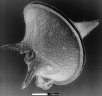
Most of the thousands of species of dinoflagellates are unicellular, yet some are colonial forms. Each species has its own specific shape. The cell wall is divided into what are called amphiesmal vesicles, which are filled with cellulose plates. The flagellar pores holding the flagella are located here in these cellulose plates. If the plates are bounded by structures called sutures, the cell wall is a theca and referred to as an armored dinoflagellate. On the other hand, a lack of a theca is known as a naked dinoflagellate.
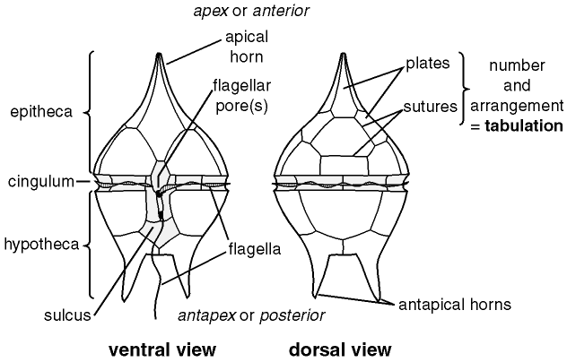
The flagellar pores usually hold two flagella into the cell wall, close to the same location. The flagella that surrounds the perimeter of the cell is called the transverse flagellum, whereas the other flagella that crosses the transverse flagellum perpendicularly is the longitudinal flagellum. The point of origin of the transverse flagellum is called the cingulum, which is like a channel that encompasses the cell except for an area known as the sulcus. A depression defines this area on the ventral surface. These two flagella together provide the forward, spinning motion of the organism, hence their name (Gr. dinos, whirling).

spiniferiteshalf.gif

Next Page